Meniscus Rupture
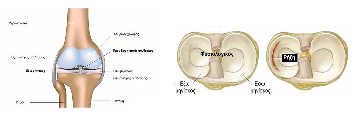
The meniscus is an integral part of the knee. The knee joint consists of three bones: The femur, tibia and patella. The patella, which is in front of the joint, provides protection. Between the femur and tibia are the menisci, two crescent-shaped structures made of cartilage that act as "shock absorbers". By their shape and composition, the two menisci (medial and lateral) contribute to the smooth functioning of the knee by smoothing the surfaces of the joint and better distributing loads.
What is a Ruptured Meniscus
Meniscus tear is one of the most common sports injuries of the knee. The type of tear depends on the morphology and location of the injury. Some of the most typical forms are bucket-handle, oblique and radial tears.
Meniscus tears can be either partial or total and can be divided into two categories: traumatic and degenerative.
• Traumatic rupture can occur at all ages but most commonly in young and active people involved in sports. Athletes after sudden and combined bending and twisting movements of the knee can rupture the meniscus.
• Degenerative rupture is found in older patients and is the result of normal aging and wear and tear of the meniscus. In older patients, the meniscus deteriorates over time and becomes more prone to injury. Even a sharp lift from a chair can be enough to cause a rupture of a degenerated meniscus.
Symptoms
Symptoms are mainly of mechanical origin and include:
• Pain on the inside or outside of the knee.
• Hydrothroat (fluid) and swelling of the joint.
• Difficulty walking, difficulty going up and down stairs and sitting or standing.
• Characteristic clicking sound, inability to fully extend the knee, while at other times the knee may lock and a feeling of instability of the knee.
The injured meniscus consists of a stable part (healthy) and an unstable part (ruptured meniscus). The unstable part gradually causes damage to the adjacent articular cartilage with the daily movements of the joint. Articular cartilage is the most valuable component of any joint and it is imperative that it is protected from possible future damage in order to prevent osteoarthritis.
After taking a complete history, the orthopaedic surgeon will examine the knee for any tenderness along the line where the meniscus is located. The presence of tenderness is consistent with a meniscus injury. Then using some useful manipulations (tests) such as the McMurray test confirms the injury.
To confirm the injury, he recommends imaging tests:
• Radiographs: Although X-rays do not depict the menisci, they can detect other associated problems, mainly of a degenerative nature, such as osteoarthritis.
• MRI: It is able to describe quite accurately an injury and in particular a rupture of a meniscus.

Treatment
The treatment depends on the size of the tear, its type, its location and the symptoms it causes.
If the symptoms do not persist and the knee is stable, the treatment is purely conservative:
• Rest: unloading the leg and avoiding daily activities for a few days.
• Ice therapy: Application of ice or cold patches for 20 minutes, several times a day.
• Elastic bandage to avoid swelling.
• Inappropriate position of the leg (higher than the rest of the body).
• Non-steroidal anti-inflammatory drugs that reduce the pain and swelling caused by the inflammatory reaction.
Thus, small chronic degenerative tears with mild symptoms can be treated conservatively with anti-inflammatory analgesic treatment, intra-articular corticosteroid injections and a specialized physiotherapy and strengthening program.
In cases where conservative treatment does not have the desired result, arthroscopy is recommended. During arthroscopy there are two possibilities: either suturing the meniscus or removing only the unstable part of the meniscus (partial meniscectomy) and normalising the remaining part. The choice of treatment depends on the age of the patient, the type of tear, the quality of the tissues and the 'age' of the lesion.
This is a surgical procedure that is performed through two small incisions (about 3 - 4 mm each) and takes 15 - 30 minutes. A partial meniscectomy (i.e. removal of the part of the meniscus that has ruptured) is usually performed. More rarely, it is sutured and reattached when certain conditions are met. Extremely rarely, a meniscus transplant or the use of an artificial substitute can be performed.
With arthroscopy, pain is reduced to a minimum, the patient's hospitalization lasts only 2 - 3 hours and the return to daily activities is usually immediate, within the next day or 2 - 3 days depending on the type of surgery (partial meniscectomy or stapling). This method is absolutely reliable and has been applied in our hospital daily for the last 25 years, giving a solution to many patients with "ruptured meniscus".
Contact the doctor to book your appointment!
The doctor will be happy to evaluate your case and recommend the optimal treatment!

